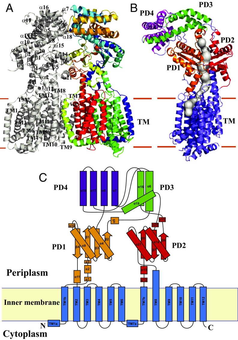
Fig. 1.
Structure of the B. mulitvorans HpnN transporter. (A) Ribbon diagram of a dimer of HpnN viewed in the membrane plane. The Right subunit of the dimer is colored using a rainbow gradient from the N terminus (blue) to the C terminus (red), whereas the Left subunit is colored gray. Overall, the HpnN dimer forms a butterfly-shaped structure. (B) Each subunit of the HpnN transport forms a channel (colored gray) spanning the outer leaflet of the inner membrane and up to the periplasmic domain. This figure depicts the Left subunit of the form I structure of the HpnN dimer. The orientation of this HpnN subunit has been rotated by 60° counterclockwise, based on the vertical C2 symmetry axis of the HpnN dimer, compared with the orientation of A. This channel was calculated using the program CAVER (loschmidt.chemi.muni.cz/caver). The transmembrane helices are colored slate. PD1–PD4 are colored orange, red, green, and magenta, respectively. (C) Secondary structural topology of the HpnN monomer. The topology was constructed based on the crystal structure of HpnN. The TM domain, PD1–PD4 are colored blue, orange, red, green, and purple, respectively.

