Fig. 5.
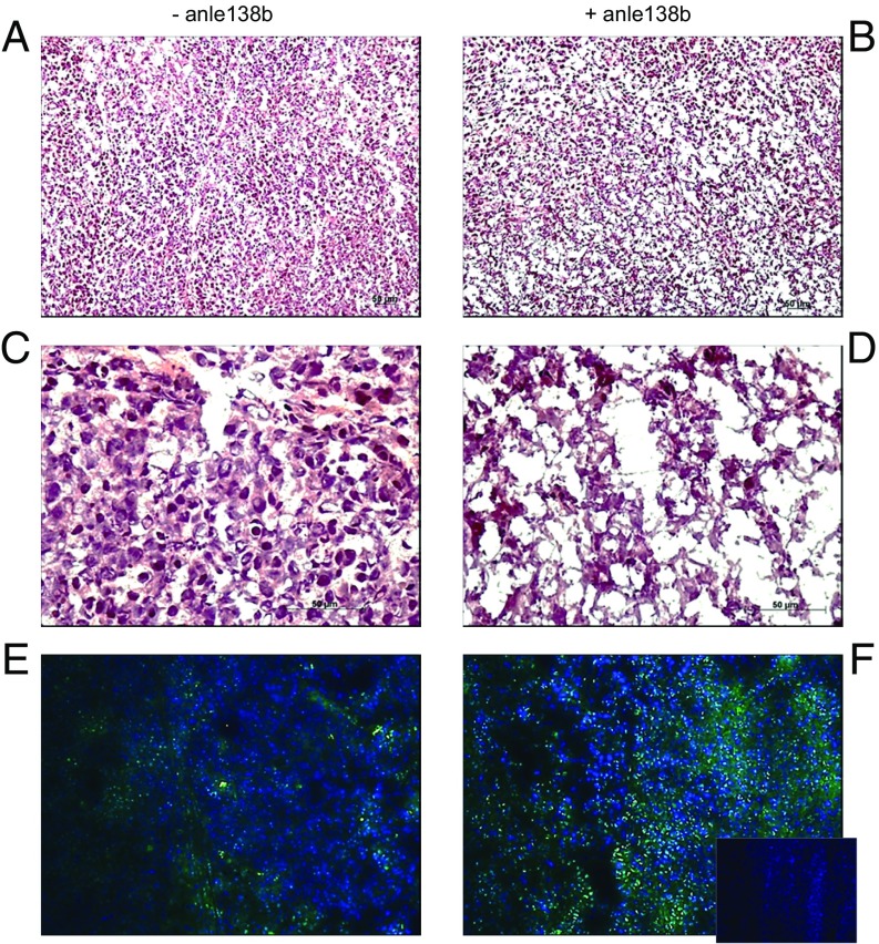
Systemic administration of anle138b to high-level α-synuclein–expressing human melanoma xenografts affects the xenografts’ morphology and autophagy. (A–D) H&E-stained tissue sections, prepared from one of the WM983-B human melanoma xenografts that had received food pellets not containing anle138b (A and C) and from one of the WM983-B human melanoma xenografts that had been resected from the animal that had received food pellets mixed with anle138b (B and D). The photographs shown in A and B were taken at 100× magnification, and in C and D at 40× magnification. (E and F) LC3 immunohistochemical staining of a tissue section (F, Inset, 100× magnification), prepared from one of the WM983-B human melanoma xenografts that had been resected from the animal that had received food pellets not containing anle138b (E) and from one of the WM983-B human melanoma xenografts that had been resected from the animal that had received food pellets mixed with anle138b (F). The anti-LC3B antibody-probed WM983-B human melanoma xenograft tissue sections (pseudocolored green) were counterstained with fluorescent DAPI (pseudocolored blue). The Inset in F shows a tissue section from the anle138b-containing WM983-B tumor, probed with Alexa-488 secondary antibody only and counterstained with fluorescent DAPI (pseudocolored blue).

