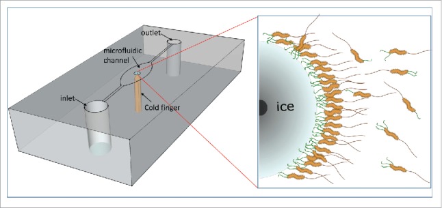
Figure 1.

M. primoryensis in the MCF device. A schematic representation of the microfluidic chip with the cold finger is presented in the left. The chip is shown upside down, and the microfluidic channel can be seen. The brass cold-finger (orange) is embedded in the middle of the chip, cooling the central part of the microfluidic well. The ice crystal grows around the tip of the cold finger in a circular shape. On the right is an enlargement of a part of the ice crystal, showing the bacteria bound to the ice surface. The black spot on the left is the tip of the cold finger. The ice-binding proteins are shown in green, and the flagella are in brown.
