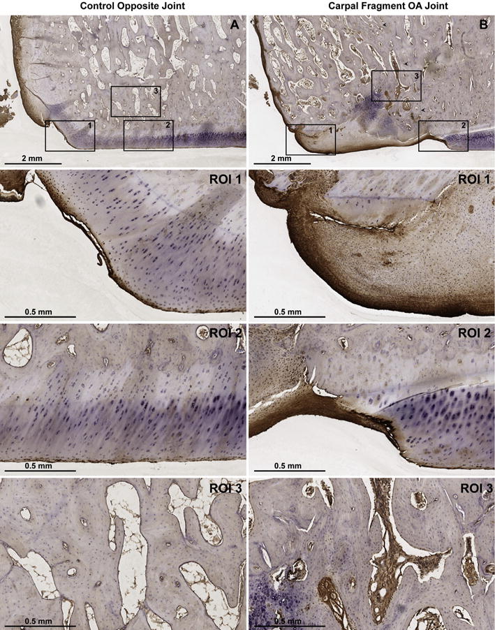
Fig. 5.

Lubricin immunostaining of the equine carpal osteochondral fragment-exercise model 70 days after fragment induction using monoclonal antibody 6a8. (A) Control and (B) fragmented articular surfaces of the distal radial carpal bone (RCB) at 1 × magnification. Arrowheads in B delineate the site of reattachment of the osteochondral fragment with the parent bone. Regions of interest (ROI) 1, 2, and 3 represent 5 × magnifications of the peripheral articular margin, the weight-bearing articular surface, and the bone within the fracture fragment (B), respectively. Lubricin immunoreaction is localized to both the surface and deeper layers of fibrocartilage (B, 1–2) as compared to the superficial layers of control hyaline cartilage (A, 1–2). Lubricin staining is also increased within the vascular channels of the RCB chip fracture fragment (B, 3) as compared to the healthy RCB (A, 3).
