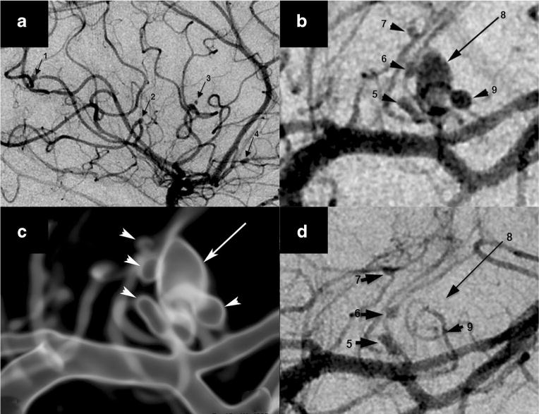
Fig. 1.

Nine aneurysms in varicella zoster virus vasculopathy. Digital subtraction angiography (DSA) demonstrated a total of nine mycotic aneurysms, four of which arose from the right middle cerebral artery (MCA) M3 segments (a, arrows 1–4) in the sylvian fissure and a cluster of left MCA lateral lenticulostriate perforator aneurysms, configured as a dominant oval aneurysm (b, long arrow 8), surrounded by four smaller lesions (b, arrowheads 5–7 and 9). A translucent rendered model from an angiographic 3D spin acquisition (c) further illustrates the complex arrangement of the left lateral lenticulostriate aneurysms. Repeat DSA after antiviral treatment and corona radiata perforator infarction showed interval thrombosis of the dominant aneurysm (d, long arrow 8) and one of the satellite lesions (d, arrowhead 9) that are no longer visible. Permission from Wolters Kluwer Health, Inc., Neurology 2014;82:2139–41
