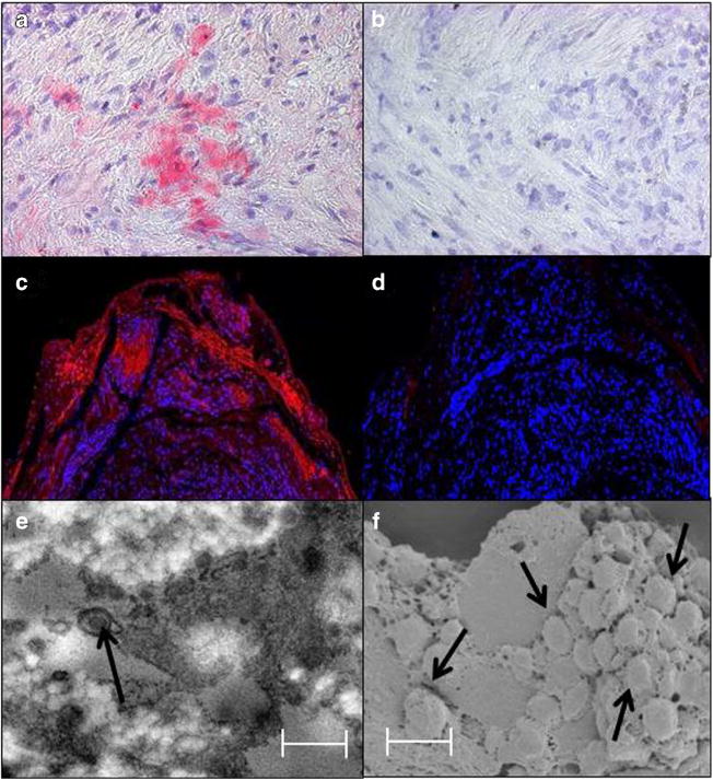
Fig. 2.

Immunofluorescence staining and ultrastructural imaging of a varicella zoster virus-infected temporal artery. Immunohistochemical staining with rabbit anti-VZV IE63 antibody revealed VZV antigen in the media (a, pink color), but not after staining with rabbit anti-HSV-1 antibody (b). Immunofluorescence staining with a different mouse anti-VZV IgG antibody than that used in panel a revealed VZV antigen in the adventitia (c, red color), but not when primary antibody was omitted (d). Transmission electron microscopy of sections adjacent to those containing VZV antigen revealed an enveloped virus particle (e, arrow), while both scanning and transmission electron microscopy of these sections showed a cluster of virus particles in the adventitia egressing through an outer cell wall (f, arrows). Virus particles appear slightly larger than 200 nm because they were sputter-coated with a gold alloy. In panels e and f, scale bars = 300 nm. Permission from Wolters Kluwer Health, Inc., Neurology 2015;84:1948–55
