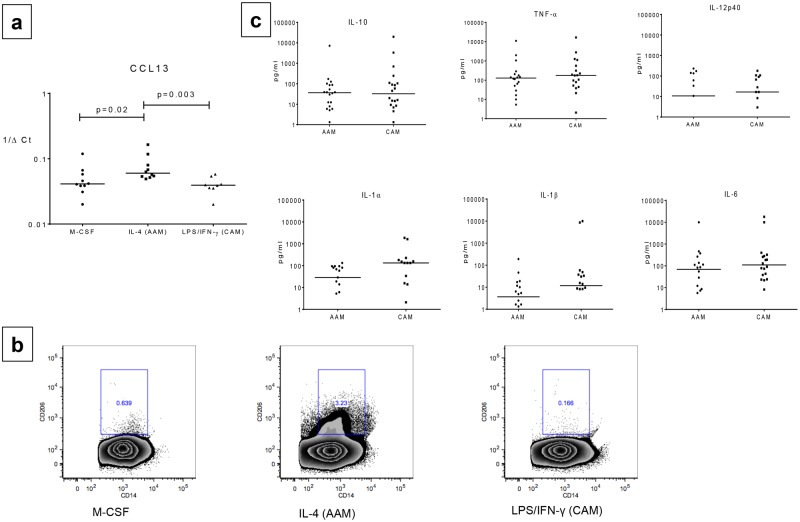
Fig 1. At baseline AAMs have increased CD206 (Mannose receptor) expression compared to CAMs.
Panel a: Relative mRNA expression of previously defined AAM-specific marker (CCL13) expressed as 1/ΔCt, was compared between alternatively activated macrophages (AAMs) and classically activated macrophages (CAMs). Individual dots representing each subject. Horizontal bars represent median. Panel b: Representative plots showing CD206 (Mannose Receptor) expression (y-axis) compared on CD14+ cells (x-axis) between M-CSF,IL-4 (AAM) and LPS/IFN-γ (CAM) by flow cytometry Panel c:total cytokine production (IL-6, IL-10, IL-12p40, IL-1α, IL-1β, and tumor necrosis factor alpha [TNF-α]) in pg/ml is compared between AAM and CAM conditions. Cells were cultured with recombinant human IL-4 (rhIL-4; 50 ng/ml) for AAMs) or with LPS (1 μg/ml)/IFN-γ (20 ng/ml) for CAMs for 48 hours. Individual dots representing each subject. Horizontal bars represent median.

