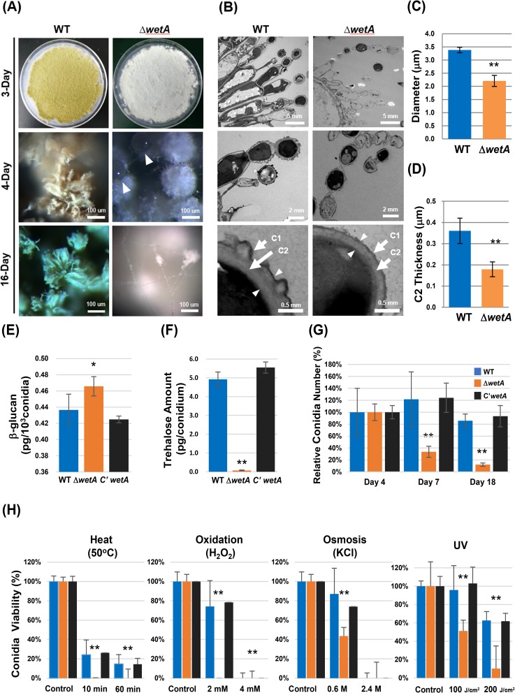
Fig 2. WetA is necessary for the proper formation of conidia in Aspergillus flavus.
(A) Phenotypes of WT (NRRL3357), ΔwetA, and C’wetA grown on solid MM at 30°C for 3, 4, 16 days after asexual induction. The white triangles indicate the liquid droplets formed on the autolyzing conidiophores of ΔwetA strain. (B) TEM images of 2-day-old conidiophores/conidia of WT and ΔwetA strains. Note: the remnant of lysed conidia formed a wet-vesicle-like structure on the top of the conidia chain, and most of the conidiophore/conidia contents were lost in the ΔwetA strain. The bottom panels show the conidia wall structures of WT and ΔwetA strains. Arrows indicate the locations of the C1 and C2 layers while the arrowheads indicate the C2 layer thickness. (C, D) The average diameter of conidia and thickness of the C2 layer of WT and ΔwetA conidia. At least seven WT and ΔwetA intact conidia from different sample slices were measured. (E, F) Quantification of conidia content (β-(1,3)-glucan (E) and trehalose (F)) of WT, ΔwetA, and C’wetA 2-day-old conidia The error bars indicate one standard deviation from the mean and the asterisks the level of significance (*, p < 0.05; **, p < 0.01). (G) The relative viability of WT, ΔwetA, and C’wetA conidia grown on solid MM at 30°C for 4, 7, 18 days after inoculation. The conidial viability at day 4 of each strain was set as 100%. ** (p < 0.01). The error bars indicate one standard deviation from the mean viability of triplicates. (H) Tolerance of WT, ΔwetA, and C’wetA 2-day-old conidia to heat (50°C), oxidative (H2O2), osmotic (KCl), and UV stresses. The control indicates untreated conidia. The viability of the untreated conidia of each strain was set as 100%. ** (p < 0.01). The error bars indicate one standard deviation from the mean viability of triplicates.

