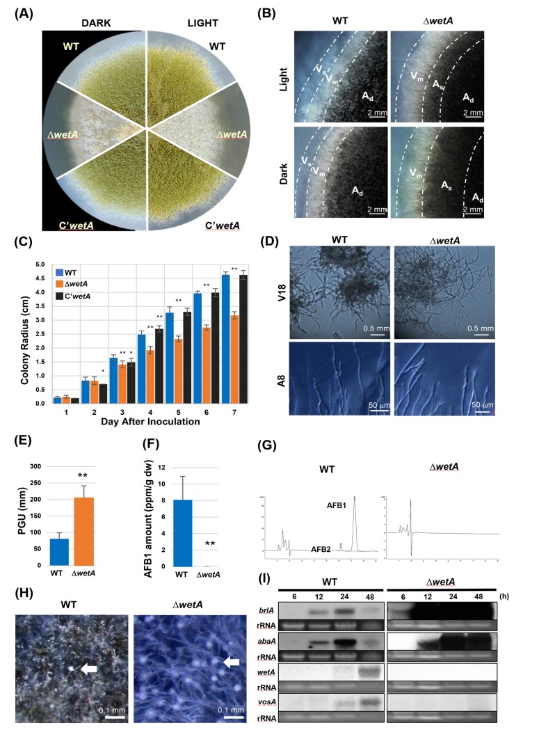
Fig 3. Multiple roles of WetA.
(A-C) WetA affects vegetative growth. (A) The colony image of WT, ΔwetA, and C’wetA strains on solid MM at 5 days after point inoculation under light and dark conditions. (B) Colony edge image of WT and ΔwetA strains under light and dark conditions. Vs: single-layer vegetative hyphae region. Vm: multi-layer vegetative region. As: sparse aerial hyphae region. Ad: dense aerial hyphae region. (C) Colony growth rates of WT, ΔwetA, and C’wetA strains after point inoculation on solid MM. The error bars indicate one standard deviation. * (p < 0.05) and ** (p < 0.01). (D, E) Hyphal branching rates of WT and ΔwetA strains. (D) Microscopy images show WetA regulates hyphal branching. Loss of wetA leads to reduced hyphal branching rate in both solid and submerged cultures. (E) Average PGU values of A8. ** (p < 0.01). The error bars indicate one standard deviation. (F, G) Aflatoxin quantification by HPLC of WT and ΔwetA submerged culture after 5-days cultivation. (F) AFB1 amount (per g dry weight) in WT and ΔwetA vegetative cells. ** (p < 0.01). The error bars indicate one standard deviation. (G) The HPLC chromatograms of AFB1 and AFB2 in the culture medium of WT and ΔwetA strains. (H) WT and ΔwetA strains were induced for asexual development and observed after 8 h incubation at 30°C on solid MM plate. The white arrows indicate conidiophores. Note: the abundant conidiophore formation in ΔwetA culture. (I) Northern blot analysis of brlA, abaA, wetA, and vosA mRNA levels in WT and ΔwetA strains at 6, 12, 24, 48 h after conidiation induction.

