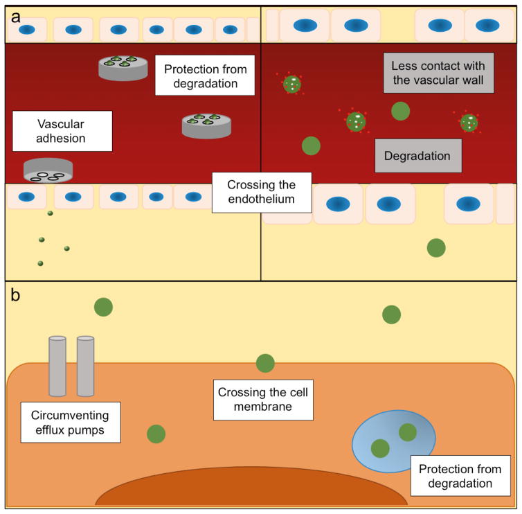
Figure 2.
Biological barriers in drug delivery. a) The MSV (left panel) and a nanodelivery system (right panel) in the circulatory system. The MSV adheres to the vascular wall, while the nanodelivery system displays less contact with the endothelium. The microparticle-component of the MSV protects nanoparticles from degradation in the circulation. The MSV forms vascular depots that gradually release nanoparticles, which can enter the interstitium through fenestrations in the vascular wall. The left and right panels have not been drawn to the same scale. b) Nanoparticles in the intracellular environment. Nanoparticles are able to cross the cell membrane, protect the payload from intracellular degradation, and circumvent drug efflux pumps.

