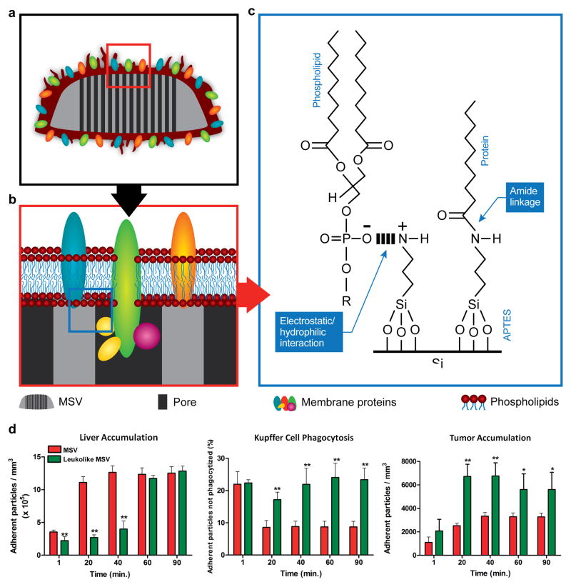
Figure 4.
Schematic of the leukolike MSV. a) The MSV was coated with cell membrane patches from leukocyte cells. b) Membrane proteins were successfully transferred onto the MSV. c) Schematic illustrating the chemical interactions between silicon and proteins/phospholipids. d) Time-dependent liver accumulation, Kupffer cell phagocytosis, and tumor accumulation of the MSV and leukolike MSV in mice. Reproduced from 57 with permission. APTES, (3-Aminopropyl)triethoxysilane.

