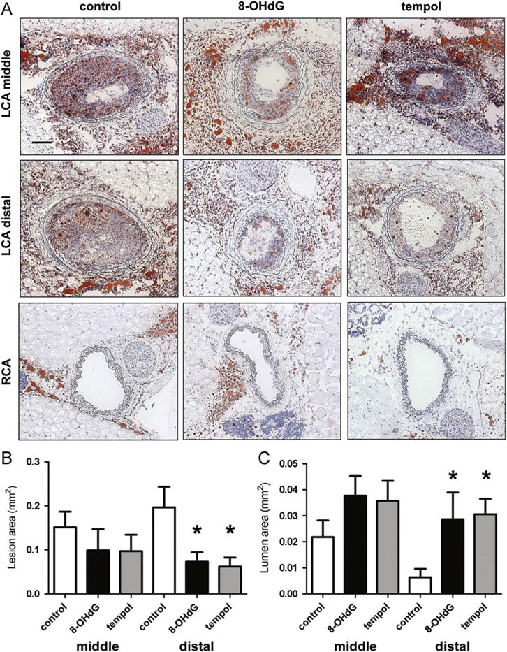
Fig. 1.

8-OHdG inhibits plaque formation in partially ligated ApoE KO mice. (A) ApoE KO mice were partially ligated and fed high-fat diet for 2 weeks. Frozen sections were stained with Oil Red O and hematoxylin. Shown are representative images of left and right carotid arteries. Scale bar; 100 μm. (B) Lesion area was calculated as the difference between internal elastic lamina area and luminal area and was quantified using Image J software. (C) Lumen area was also quantified. Values are means±SE of 6 mice per group. *P<0.05 vs. control distal left carotid artery (LCA). (For interpretation of the references to color in this figure legend, the reader is referred to the web version of this article.)
