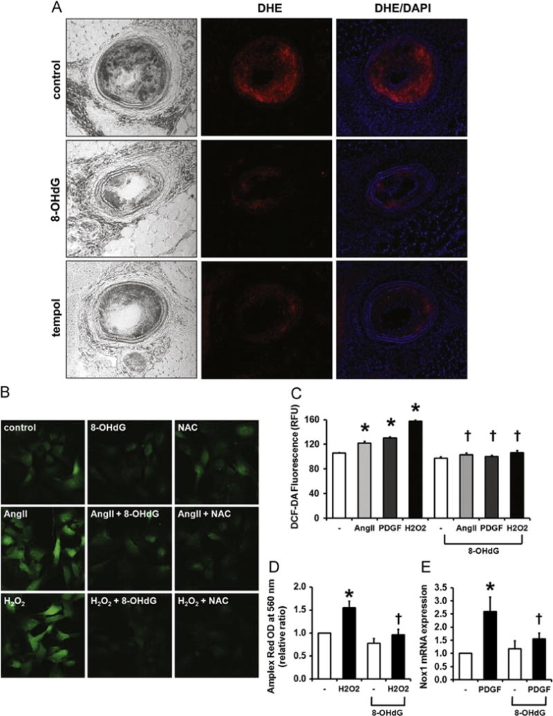
Fig. 2.

8-OHdG inhibits ROS formation in ApoE KO mice and cultured VSMCs. (A) Frozen sections of LCAs stained with DHE show decreased superoxide production in 8-OHdG treated aortas. DAPI was used to visualize nuclei. (B) Representative image of DCF-DA stained VSMCs. VSMCs were pretreated with 8-OHdG (100 μg/mL) or N-acetyl cysteine (NAC, 5 mM) for 1 h and stimulated with Ang II (1 μm) or H2O2 (1 mM) for 30 min. After stimulation, DCF-DA (5 μm) was loaded for 20 min, then fixed and mounted. (C) DCF-DA fluorescence was quantified by fluorometer in Ang II (1 μm), PDGF-BB (100 ng/mL), or H2O2 (1 mM) treated VSMCs. 8-OHdG (100 μg/mL) was pretreated for 1 h. (D) Secreted ROS was detected using Amplex Red. VSMCs were pretreated with 8-OHdG (100 μg/mL) for 1 h and stimulated with H2O2 (1 mM) 20 min. To remove the H2O2 stimulated, the cells were changed to fresh media and incubated for 30 min. Amplex Red (50 μm) was added to the medium the absorbance was detected at 560 nm. (E) Relative mRNA expression of Nox 1 in PDGF-BB treated VSMCs. 8-OHdG (100 μg/mL) was pretreated for 1 h and stimulated with PDGF-BB (10 ng/mL) for 24 h. Values are means±SE of 4 experiments. *P<0.05 vs. control, †P<0.05 vs. Ang II or PDGF-BB treated cells. DHE; dihydroethidium. (For interpretation of the references to color in this figure legend, the reader is referred to the web version of this article.)
