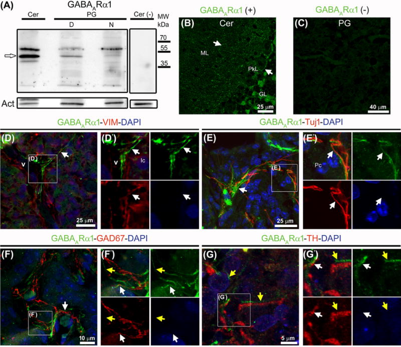
Fig. 2.

Western blot and immunohistochemistry reveal the ionotropic receptor subunit GABAARα1 in the adult rat pineal gland. (A) A band around 51 kDa (white arrow) seen in whole extracts from cerebellum (Cer) and pineal gland (PG) pools at midday (D) and midnight (N). Omission of the primary antibody and actin (Act) served as negative and loading controls, respectively. (B–G′) Immunolabeling for GABAARα1 (green), vimentin (VIM, red), Tuj1 (red), GAD67 (red), and tyrosine hydroxylase (TH) (red); nuclei were counterstained with DAPI (blue). (B) Punctate pattern of the ionotropic subunit in the cytoplasm, surface and dendritic tree of Purkinje cells (PkL, white arrows). (C) Pineal gland without primary antibody. (D–G′) In pineal gland sections a specific signal for GABAARα1 is seen as extensions often interlocked with or parallel to VIM-positive blood vessels (v) and in a more discontinuous way, to sympathetic and GABAergic nerve fibers (white arrows). A signal not associated directly with pineal gland innervation is marked with yellow arrows. (D′, E′, F′, G′) Higher magnifications of the insets shown in D–G. Abbreviations: GL, cerebellar granular layer; Ic, interstitial cells; ML, cerebellar molecular layer; MW, molecular weight; Pc, pinealocytes. (B) 1.1× magnification of a 60× image. (D, E) 2× digital zooms of 60× images. (D′, E′) 2× enlargements of the insets shown in D and E. (F) 1.2× magnification of a 60× image. (G) 4× digital zoom of a 100× image. (F′, G′) 2× and 1.5× enlargements of the insets shown in F and G, respectively.
