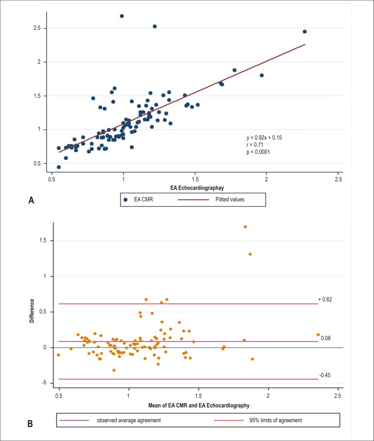
Figure 4.
Results obtained using cardiac magnetic resonance (CMR) three-dimensional volume-curve and echocardiography Doppler mitral valve inflow. The ratio between the early peak filling (E) and atrial peak filling rate (A) using velocity (cm/s) by echocardiography and flow (mL/s) by CMR. (A) Linear regression and Pearson's correlation; (B) Bland-Altman analysis.

