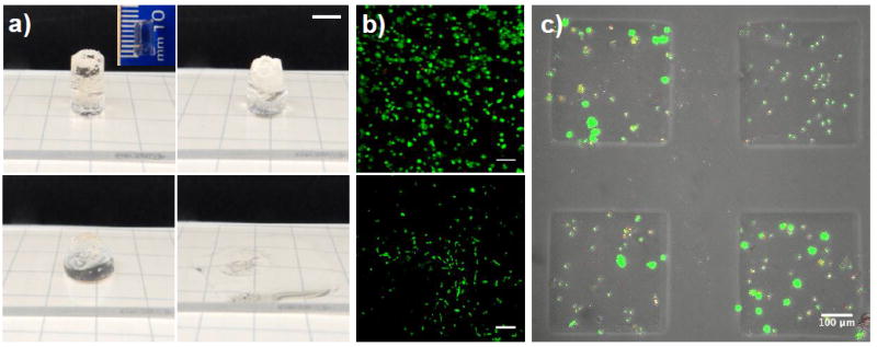
Figure 4. Allyl sulfide based photodegradable hydrogels can be used for large scale erosion and cytocompatible cell release.

a) A 1 cm thick hydrogel containing 4 mM LAP and 25 mM mPEG-SH was irradiated with 10 mW cm-2 365 nm light, and completed eroded over the course of approximately 1 min. In this optically thick gel, 87 % of the incident light is attenuated through the sample (i.e., only 13% of the light reaches the bottom of the sample). Macroscopic images of the gel are shown at 0 s, 20 s, 44 s and 74 s (left to right, top to bottom). Scale bar 5 mm. b) Cells that were both encapsulated for 24 h and subsequently released remained highly viable. Top panel: hMSCs 24 h after encapsulation are 90% viable. Bottom panel: released cells remain viable and spread on glass coverslips over 24 h. Cells were stained with calcein AM (green, live) and ethidium homodimer (red, dead). Scale bars: 100 μm (top panel) and 300 μm (bottom panel). c) A 150 μm thick cell-laden hydrogel was selectively exposed to light (365 nm at 10 mW cm-2) for 1 min through a photomask to induce spatially controlled photodegradation and release a subset of the encapsulated cells. Cells remaining in hydrogel monoliths stained with with calcein AM (green) and ethidium homodimer (red). Scale bar = 100 μm.
