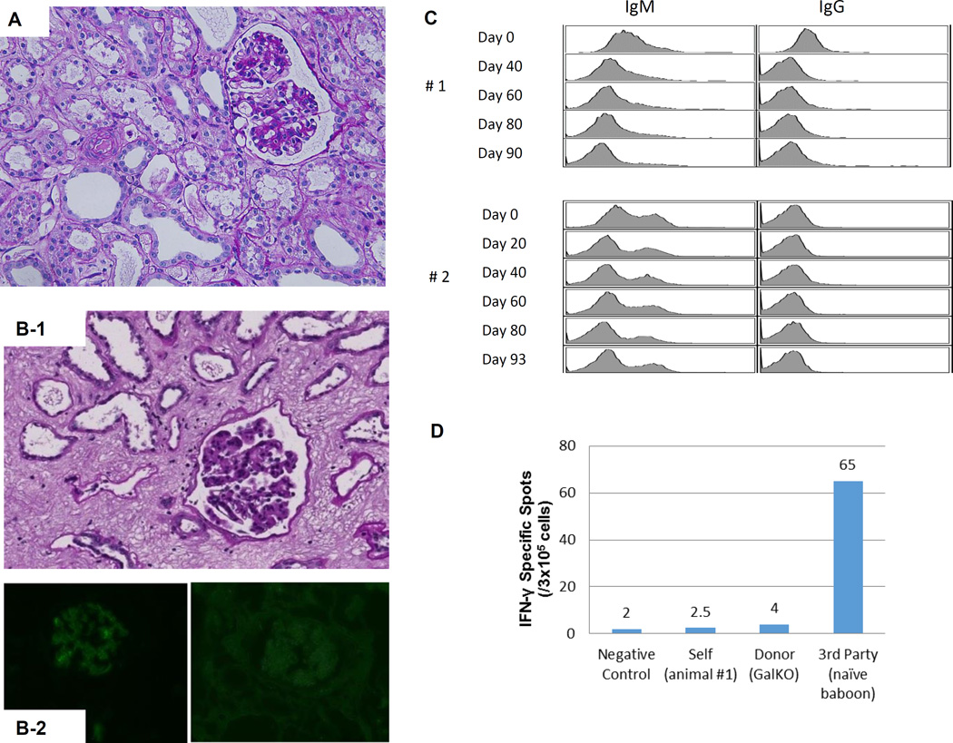
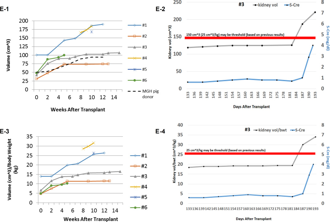
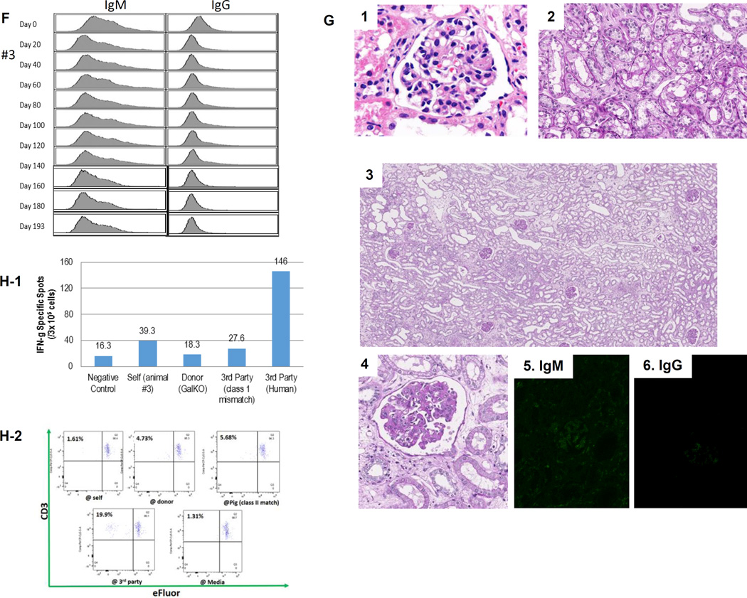
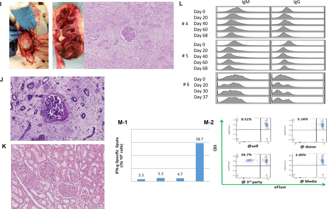
Figure 1.
(A) Periodic acid-Schiff (PAS) staining of a kidney graft for baboon #2 on day 93. No sign of rejection or interstitial fibrosis. (B-1) PAS staining of a kidney graft for baboon #1 on day 90 showed interstitial fibrosis and tubular atrophy but no evidence of glomerulitis, peritubular capillaritis or a mononuclear cell infiltration. (B-2) Immunofluorescence staining for baboon #1 on POD 90. IgM staining showed mild mesangial deposition (left) while IgG deposition wasn’t seen (right). (C) Anti-non-Gal natural antibody for baboons #1 and #2 assessed by flow-cytometry demonstrating neither elicit anti-pig IgM nor IgG developed.(D) ELISPOT assay for IFN-γ at the time of euthanasia (POD 90) for baboon #1. Many spots could be seen in the wells stimulated by 3rd party while only a small number of spots against GalTKO pig cells similar to self were seen. (E-1) Kidney volume (cm3) of all xenoTK Tx recipients against weeks after Tx (solid lines). The growth of the TKs (solid lines) was similar to what was observed in a remaining kidney of a miniature swine donor who was kept alive after kidney donation in order to measure autologous kidney growth (dashed line).(E-2) Kidney volume and serum Cre levels between 19 and 27 weeks after Tx for baboon #3. (E-3) Ratio of kidney volume (cm3) / recipient’s body weight (kg) against weeks after Tx. (E-4) Ratio of kidney volume (cm3) / recipient’s body weight (kg) and serum Cre levels for baboon #3 between 19 and 27 weeks after Tx.(F) Anti-non-Gal natural antibody for baboon #3 assessed by flow-cytometry demonstrating neither elicit anti-pig IgM nor IgG developed.(G-1 and 2) The graft biopsy at POD 121 from baboon #3 showed no interstitial edema and normal appearance of glomeruli. (G-3 and 4) Biopsy at POD 193 from baboon #3 showed well preserved glomeruli with no glomerulitis, no endoarteritis, and no apparent interstitial cell infiltration. However, widening of interstitial area was noted with interstitial edema. (G-5 and 6) Neither IgM nor IgG deposition was observed in the excised grafts from baboon #3. (H-1 and 2) ELISPOT assay for IFN-γ and MLR at the time of euthanasia for baboon #3 demonstrated that animal was anti-pig specific unresponsive. (I) Necropsy sample of kidney graft (left) and PAS staining of kidney graft for baboon #4 on POD 68 (right). Glomerular capillaries were relatively preserved with diffuse tubular necrosis and absence of thrombi in the small interlobular arteries. (J) PAS staining of a kidney graft for baboon #5 on day 68 showed interstitial fibrosis and tubular atrophy but glomerular capillaries were relatively preserved. (K) Kidney graft for baboon #6 on day 37 showed no sign of rejection. (L) Anti-non-Gal natural antibody for baboon #4, #5 and #6 assessed by FACS demonstrating neither elicit anti-pig IgM nor IgG developed. (M-1) INF-γ ELISPOT assay for baboon #4 at the end of study (POD68). (M-2) MLR for baboon #4 on day 59.


