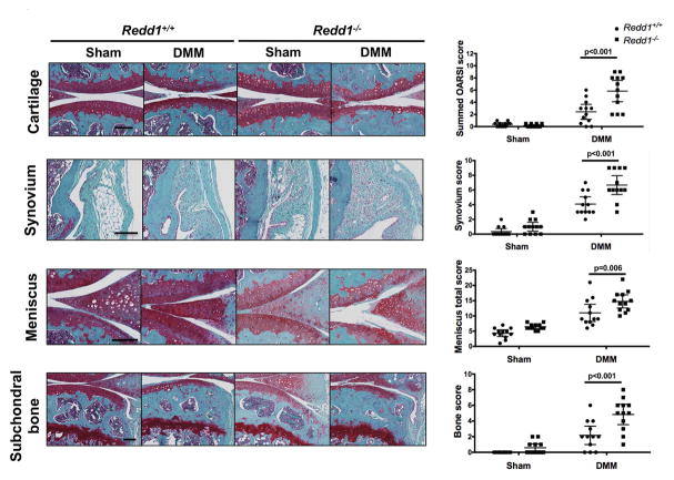
Figure 1.
Increased severity of experimental OA in Redd1−/− mice. A) Representative images of knees showing histological changes in the medial articular cartilage, synovium, meniscus and subchondral bone from 6.5-month-old Redd1+/+ and Redd1−/− mice (n=12 per genotype) subjected to DMM or sham procedure. Right panels show quantification of the histological scores for each joint tissue. All values are mean ± SD. Scale bar = 100 μm.

