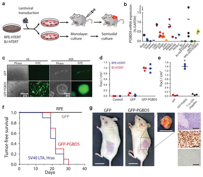
Fig. 3. Ectopic expression of PGBD5 in human cells leads to oncogenic transformation both in vitro and in vivo.

(a) Schematic for testing transforming activity of PGBD5. (b) Relative PGBD5 mRNA expression measured by quantitative RT-PCR in normal mouse tissues (brain, liver, spleen and kidney), as compared to human tumor cell lines (rhabdoid G401, neuroblastoma LAN1 and SK-N-FI, medulloblastoma UW-228 cells), primary human rhabdoid tumors (PAKHTL, PARRCL, PASYNF, PATBLF), and BJ and RPE cells stably transduced with GFP-PGBD5 and GFP. Error bars represent standard deviations of 3 independent measurements. (c) Representative images of GFP-PGBD5-transduced RPE cells grown in semisolid media after 10 days of culture, as compared to control GFP-transduced cells. (d) Number of refractile foci formed in monolayer cultures of RPE and BJ cells expressing GFP-PGBD5 or GFP, as compared to non-transduced cells (p = 3.6 × 10⁻⁵ and 3.9 × 10⁻⁴ for GFP-PGBD5 vs. GFP for BJ and RPE cells, respectively). (e) Expression of T. ni GFP-PiggyBac does not lead to the formation of anchorage independent foci in monolayer culture (* p = 3.49 × 10⁻⁵ for GFP-PGBD5 vs. T. ni GFP-PiggyBac). Error bars represent standard deviations of 3 independent experiments. (f) Kaplan-Meier analysis of tumor-free survival of mice with subcutaneous xenografts of RPE cells expressing GFP-PGBD5 or GFP control, as compared to non-transduced cells or cells expressing SV40 large T antigen (LTA) and HRAS (n = 10 mice per group, p < 0.0001 by log-rank test). (g) Representative photographs (from left) of mice with shaved flank harboring RPE xenografts (scale bar = 1 cm). Tumor excised from mouse harboring GFP-PGBD5 expressing tumor (scale bar = 1 cm). Photomicrograph of GFP-PGBD5 expressing tumor (top to bottom: hematoxylin and eosin stain, vimentin, and cytokeratin, scale bar = 1 mm).
