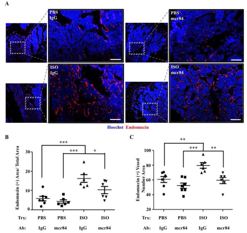
Figure 5. Blocking the Interaction Between Vegf-a and Vegfr2 Reduces the Increase in Bone Vascular Density Caused by ISO Administration.

A) Representative 10× and 20× confocal images of hind limb paraffin sections from mice treated with PBS or ISO, and with either mcr84 or IgG2a control antibody for 6 wks, stained for endomucin (IF, red). Hoechst= Blue. Bar: 100 μm. B) Quantification of the Endomucin-positive area/Total Area. C) Quantification of the number of Endomucin (+) Vessels/Area (*P=<.05, **P=<.01, ***P=<.001, N=6–7).
