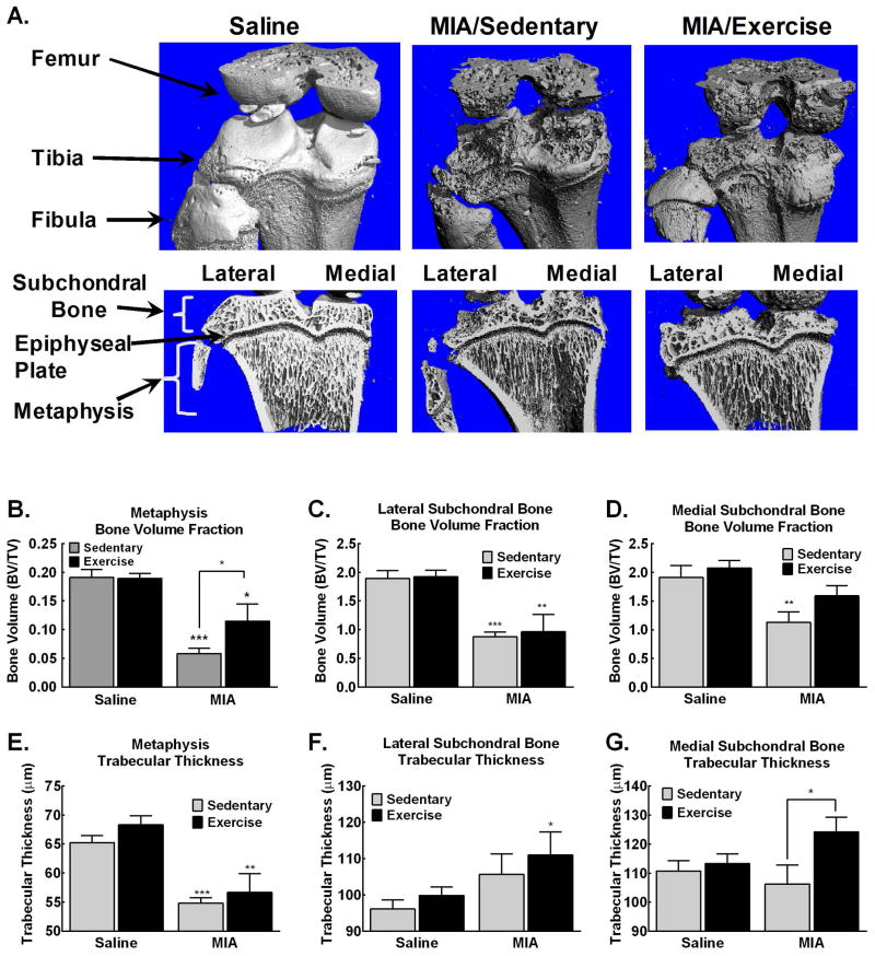
Figure 5. MIA-induced pathological changes in the subchondral and metaphysis are blocked by treadmill exercise.
A. Representative images demonstrate MIA-induced bone remodeling of the exterior bone, with development of osteophytes and bone deformities. Trabecular bone loss is observed within subchondral bone and metaphysis. B. Significant reduction in bone volume is observed in the medial subchondral bone of MIA treated sedentary, but not MIA treated exercise rats. C. Significant reduction in bone volume is observed in the lateral subchondral bone of MIA treated sedentary and exercise treated rats. D. Diminished bone volume is observed in the trabecular region of the metaphysis in the MIA treated sedentary rats that is attenuated in the MIA treated rats that underwent treadmill exercise. E. Significant reduction of trabecular bone thickness is observed in the metaphysis of in MIA treated rats. F. Increased trabecular thickness is observed in the lateral subchondral bone of MIA treated rats. G. Increased trabecular thickness within the medial subchondral bone is observed in MIA treated rats that underwent exercise across 4 weeks. *p<0.05, **p<0.01, ***p<0.001 vs saline sedentary.

