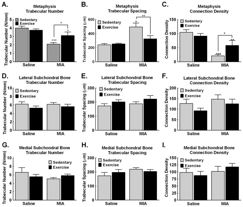
Figure 6. MicroCT analysis of the tibia demonstrates that MIA-induced pathological changes in the subchondral and metaphysis are blocked by treadmill exercise.
A. Analysis of the number of trabeculae within the metaphysis demonstrates decreased trabeculae in MIA treated sedentary rats. Exercise blocked the MIA-induced decrease in trabecular number, but the number was still significantly lower compared to saline sedentary control rats. B. Analysis of trabecular spacing demonstrates increased space between trabecular bone within the metaphysis in the sedentary MIA treated rats. Exercise blocked the MIA-induced increase in trabecular spacing, with trabecular spacing similar to saline sedentary control rats. C. MIA reduced connection density of trabecular bone within the metaphysis. Exercise attenuated the MIA induced reduction in connection density of trabecular bone. D–F. MIA and exercise both failed to alter the number (D), spacing (E) or connection density (F) of the trabecular bone within the lateral subchondral bone. G–I. MIA and exercise both failed to alter the number (G), spacing (H) or connection density (I) of the trabecular bone within the medial subchondral bone. Graphs represent mean ± SEM. *p<0.05, **p<0.01, ***p<0.001 vs saline sedentary.

