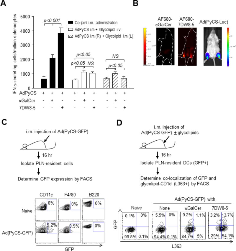
Figure 1.

The potent 7DW8-5 adjuvant effect is dependent on the route of its administration. (A) Groups of BALB/c mice were administered i.m. (anterior tibialis muscles) with 5×10⁸ pfu AdPyCS alone or co-jointly with 1 μg αGalCer or 7DW8-5. The solid column represents co-joint i.m. administration of the vaccine and respective glycolipid; the unfilled column represents i.m. administration of the vaccine, immediately followed i.v. administration of each glycolipid; the striped column represents i.m. administration of the vaccine and glycolipid in opposite legs. Twelve days later, splenocytes were harvested, and the relative number of PyCS-specific CD8+ T cells was determined with IFN-γ ELISpot assay. The results are expressed as mean ± S.D of four mice in each group. (B) BALB/c mice (n=4/group) were injected i.m. (anterior tibialis muscles) with 5 μg AF680-αGalCer or AF680-7DW8-5, and 12 hours later, images were collected using Lumina IVIS. Images from one representative mouse are shown. Another group of four BALB/c mice were injected i.m. with 5 × 10⁹ v.p. of Ad(PyCS-Luc). Twelve hr later, mice were i.p. injected with 200 μL of 15 mg/mL D-luciferin, and fluorescent signal of luciferin was collected with Lumina IVIS. One representative image is shown. (C) BALB/c mice (n=4/group) were injected i.m. with 5×10⁹ v.p. of Ad(PyCS-GFP). Sixteen hr after immunization, lymphocytes were isolated from PLNs, and GFP expression in DCs, macrophages and B cells were determined by flow cytometry. Naïve mice were used as a negative control. Histograms from one representative mouse in each group are shown. (D) Sixteen hr after Ad(PyCS-GFP) and glycolipid immunization later, PLNs were isolated and GFP-expressing DCs and CD1d bound glycolipids were determined by anti-L363 antibody. DCs collected from naïve mice were a negative control. Histograms from one representative mouse in each group are shown.
