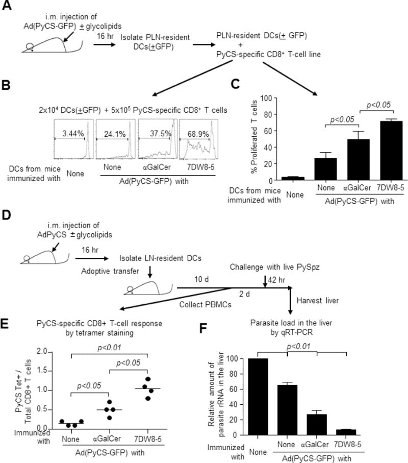
Figure 3.

(A) BALB/c mice (n=20/group) were immunized i.m. with 5×10⁹ v.p. of Ad(PyCS-GFP) alone or together with 1 μg of each glycolipid. Sixteen hr later, GFP+ DCs were sorted, and 2 × 10⁴ GFP+ DCs were co-cultured with 5×10⁵ CFSE-labeled, PyCS-specific CD8+ T cells in triplicate. Four days later, CFSE fluorescence was determined by flow cytometry. (B) Histograms from one representative experiment and (C) the mean ± S.D. of the triplicate experiments are shown. (D) BALB/c mice (n=20/group) were immunized i.m. with 5 × 10⁹ v.p. of AdPyCS alone or together with 1 μg of each glycolipid. Sixteen hr later, PLNs-resident DCs were isolated and adoptively transferred i.v. to naïve BALB/c mice at 7.5 × 10⁵ cells/mouse. (E) Ten days later, blood was collected and the percentage of PyCS-specific CD8+ T cells present among CD8+ T cells was determined by a flow cytometric analysis using SYVPSAEQI-loaded H-2Kd tetramer. (F) Two days later the same transferred mice, as well as naïve BALB/c mice were challenged with 2 × 10⁴ live PySpz by i.v. and 42 hr later and the parasite burden in the liver was determined by quantifying the amount of parasite-specific rRNA by a real-time qRT-PCR.
