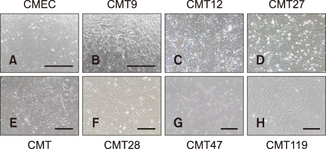
Fig. 1. Canine mammary epithelial cells (CMEC) and canine mammary tumor (CMT) cells. Phase contrast images of live CMT cells in culture were taken at 50–100% confluence in culture media by using a photomicroscope. The CMT primary cell image shows a representative colony of mammary carcinoma cells surrounded by a field of mammary fibroblasts prior to high-speed cell sorting on a MoFlo flow cytometer and cell sorter (Beckman Instruments). Magnifications for CMT12 and CMT27 cells were the same as CMT primary cell cultures. Representative images are shown. Scale bars = 1,000 µm (A and B), 400 µm (E and G), 200 µm (F and H).

