. 2017 Jun 22;18(2):149–158. doi: 10.4142/jvs.2017.18.2.149

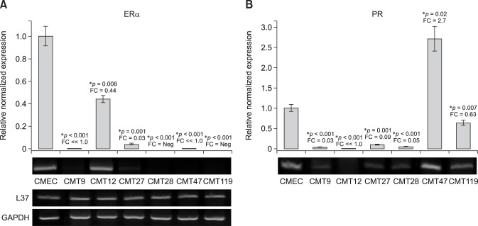
Fig. 2. Expression of canine estrogen receptor α and progesterone receptor genes in canine mammary tumor (CMT) Cells. Quantitative reverse transcriptase polymerase chain reaction (QrtPCR) assays were developed to amplify discreetly each canine receptor mRNA in order to assess expression levels for each receptor in total cell RNA isolated from CMT cell lines CMT9, CMT12, CMT27, CMT28, CMT47, and CMT119. Agarose gel electrophoresis of semi-quantitative rtPCR for each amplicon is also shown to validate the QrtPCR values. (A) mRNA levels of estrogen receptor α (ER1) assessed by QrtPCR assay and normalized, for recovery and assay artifact, to expression levels of GAPDH and ribosomal protein L37 transcripts (L37). Normalized values were then expressed as a relative fold-function of the expression levels observed in normal canine mammary epithelial cells (CMEC), which were set to a value of 1 in the same assay. (B) mRNA levels of progesterone receptor (PR) were assessed by QrtPCR assay and calculated as described for ER1 mRNA expression. Agarose gels showing semi-quantitative levels of ERα and PR transcripts as well as GAPDH and L37 transcripts, which served as positive controls used for normalization, are also shown as positive controls to demonstrate the consistency of expression across samples. Significant differences, at the confidence levels (p values) noted, between CMEC and CMT expression levels are noted. An asterisk indicates significance at p≤0.05 or 0.01 as noted. FC, fold-change relative to control cells (CMEC); Neg, not expressed/detected in semi-quantitative or QrtPCR assays.

Fig. 2