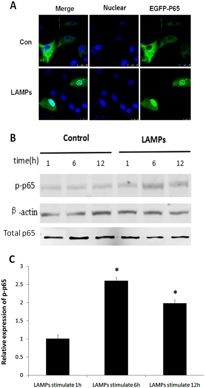
Figure 2.

NF-κB is activated following LAMP stimulation. (A) EBL cells were transfected with an EGFP-p65 expression plasmid (pEGFP-p65) for 24 h and then stimulated with 1.0 μg/mL LAMPs for 6 h. Nuclei were counterstained with 1 μg/mL DAPI, and NF-κB nuclear translocation was observed using a laser-scanning confocal microscope. (B) EBL cells were stimulated for the indicated times with 1.0 μg/mL LAMPs, and Western blot analyses were performed with antibodies specific for bovine p65 and p-p65. β-Actin was used as a loading control. (C) Band densitometry results from the western blot shown in (B). β-Actin was used for normalization. *p < 0.05 (Student’s t-test).
