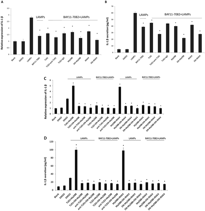
Figure 7.

Mmm-derived LAMPs activate IL-1β production through the NF-κB pathway via TLR2, MyD88, and IRAK4. (A and B) EBL cells were transfected with 0.2 μg/mL pHA-TLR2, 0.2 μg/mL pHA-MyD88, 0.2 μg/mL pHA-DN-MyD88, 0.2 μg/mL pHA-IRAK4, or 0.2 μg/mL pHA-DN-IRAK4. After 24 h, the cells were incubated with 10 μg/mL of an anti-TLR2 mAb or an isotype-matched human IgG for 30 min, stimulated with 1.0 μg/mL LAMPs, and treated with the NF-κB inhibitor BAY11-7082 (10 μM) or DMSO (control) for 6 h. The cells and supernatants were then respectively harvested separately and analyzed by (A) real-time PCR and (B) ELISA. Data are presented as the mean and standard deviation of three assays. *p < 0.05 compared with DMSO-treated, LAMP-stimulated cells. (C and D) EBL cells were transiently cotransfected with the indicated constructs (0.2 μg/mL pHA-TLR2, pHA-MyD88, pHA-IRAK4, pHA-DN-MyD88, and pHA-DN-IRAK4). After 24 h, the cells were treated or not with 10 μg/mL anti-TLR2 IgG mAb for 30 min, stimulated with 1.0 μg/mL LAMPs, and treated with an NF-kB inhibitor (10 µM) or DMSO (vehicle control) in the absence of serum for 6 h. The cells and supernatants were then respectively harvested and analyzed by (C) real-time PCR and (D) ELISA. Data are presented as the mean and standard deviation of three assays. *p < 0.05 compared with DMSO-treated, LAMP-stimulated cells.
