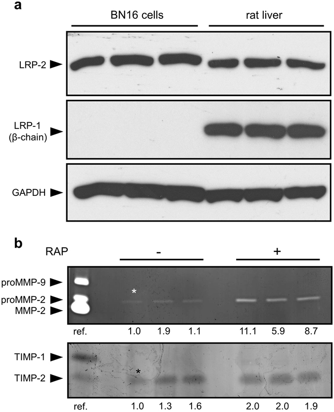
Figure 1.

Expression of functional megalin/LRP-2 but not LRP-1 in BN16 cells. (a) Expression of megalin/LRP-2 and LRP-1 was evaluated by western blot analysis in BN16 cell extracts (50 μg protein). As positive control, receptor expression was determined in tissue extracts (50 μg protein) from rat liver40. Images were cropped for presentation. Additional data without cropping are presented in Supplementary Fig. 1. (b) Analysis of media conditioned by BN16 cells in the presence of the LRP competitor, RAP. BN16 cells were cultured for 24 h in serum-free DMEM in the absence or presence of 1 μM RAP. Conditioned media were then collected, and total cell protein was measured using bicinchoninic acid microassay. Top panel, gelatin zymogram of medium conditioned by the equivalent of 5 μg of cell protein. Bottom panel, reverse gelatin zymogram of medium conditioned by the equivalent of 10 μg of cell protein. Representative results of three independent experiments. Reference (ref.) corresponds to medium conditioned by mouse calvarium, as previously reported41. Values under the gels indicate the fold-increase by comparison with the first non-treated sample (*). Band area and intensity were measured by using IMAGEJ image analysis software.
