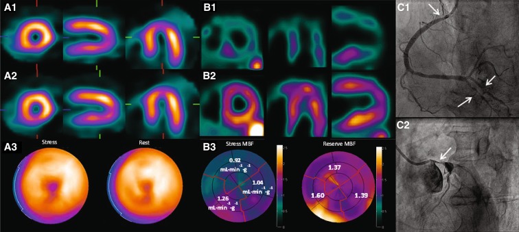
Fig. 3.
99mTc-Tetrofosmin SPECT (A), H2 15O PET (B) and invasive coronary angiography (C) images of a 73-year-old male with chest pain. Short and long axis images during stress (A1) and rest (A2) show a homogenous tracer distribution indicating a normal perfusion. Stress and rest polarmaps (A3) display the same normal perfusion pattern. PET derived short and long axis images show an extensive attenuated perfusion pattern during stress (B1) as compared to rest (B2). Quantitative MBF values provided in the polarmaps (B3) indicate ‘balanced ischemia’ with impaired hyperemic MBF (<2.30 mL min−1 g−1) and flow reserve (<2.50) values in each vascular territory. Invasive coronary angiography confirmed the diagnosis with multi vessel disease located at the proximal and distal right coronary artery (C1) and left main coronary artery (C2) as indicated with arrows

