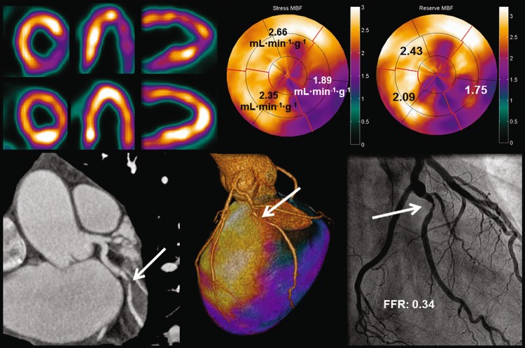
Fig. 5.
Case example of a 46-year-old male with typical anginal chest pain. PET showed an inferolateral perfusion defect with an abnormal hyperemic perfusion of 1.89 mL min−1 g−1 and a myocardial flow reserve of 1.75. CCTA displayed an obstructive soft plaque located in the obtuse marginal branch. Fused PET and CCTA images revealed a perfusion defect downstream from the coronary stenosis. Invasive coronary angiography showed angiographic significant luminal narrowing of the obtuse marginal branch (1-vessel disease) with an FFR of 0.34

