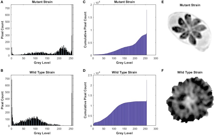
Figure 3.
Gray-level histogram for mutant and wild-type Toxoplasma gondii strains. (A,B) show the number of pixels in each gray level (0–256) of the images of the parasitophorous vacuole (PV) with 0 signifying black and 256 signifying white. (C,D) show the cumulative pixel count for the same images, which are shown in (E,F). This shows that PV produced by wild-type strain had a significantly larger fraction of darker pixels, which represents the large number of the growing intra-vacuolar parasites, whereas the PV of mutant strain had significantly lower quantity of the intra-vacuolar parasites, as shown by a greater proportion of the ligher pixels.

