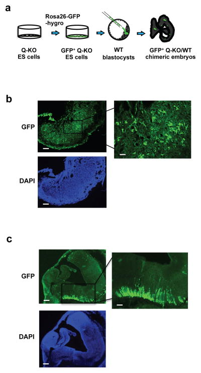
Figure 5. Contribution of Q-KO ES cells to the developing embryo.
a, Strategy used to gauge the contribution of Q-KO (GFP+) ES cells to different compartments of the developing embryos. b, Strong contribution of Q-KO (GFP+) ES cells to the embryonic part of placenta. Shown is a section of a placenta from a chimeric embryo, stained for GFP. Scale bar, 100 μm. Right panel shows higher magnification of the boxed area, scale bar, 400 μm. Lower panel represents DAPI staining (to visualize cells). c, A section of embryonic brain from a chimeric embryo stained for GFP. Scale bar, 100 μm. Right panel shows higher magnification of the boxed area, scale bar, 400 μm. Lower panel represents DAPI staining. Three Q-KO and one Ctrl GFP+ ES cell lines were injected, 10 Q-KO and 3 Ctrl embryos were analyzed. The imagines are representative of 10 Q-KO chimeric embryos.

