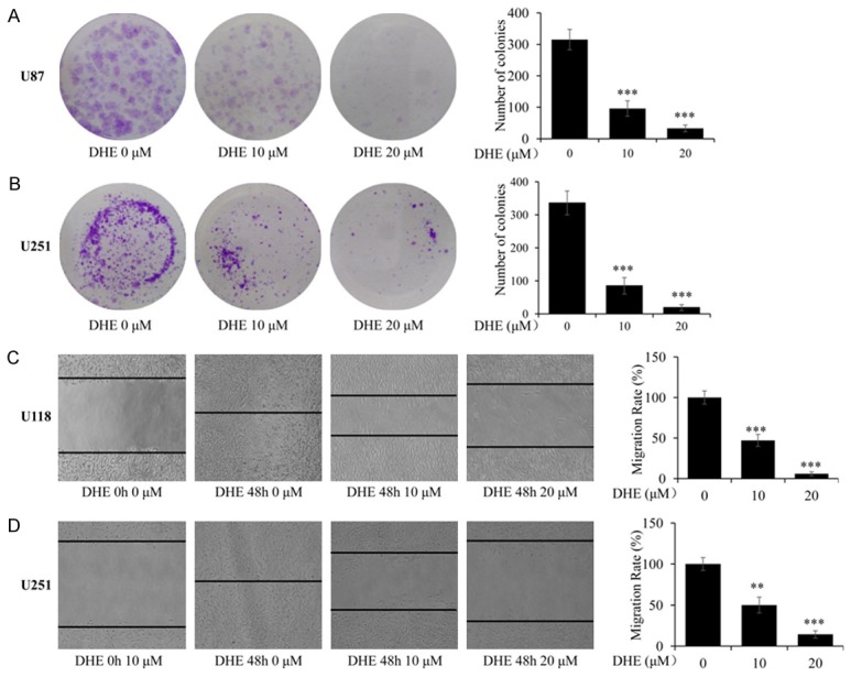
Figure 2.

DHE suppressed clonogenic and migratory ability of glioblastoma cells. (A, B) U87 and U251 cells were treated with different concentrations of DHE (0, 10 and 20 μM) for 3 h, and then cultured for an additional 15 days. Representative images of the U87 (A) and U251 (B) cells were captured using a microscope fitted with digital camera (magnification, ×100). And the colonies number of the U87 and U251 cells was calculated. (C, D) Cells were treated with DHE (0, 10 and 20 μM) for 48 h after scratch. Representative images of the U118 (C) and U251 (D) cells were captured at 0 h and 48 h using a microscope fitted with digital camera (magnification, ×100). And the migration rate of the U118 and U251 cells was calculated. The data are presented as the mean ± SD of three independent experiments. (*P<0.05, **P<0.01 and ***P<0.001 as compared with the control group).
