Figure 1.
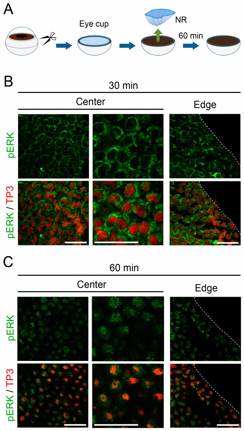
Retinectomy is a causal event for MEK-ERK signal reinforcement in retinal pigment epithelium (RPE) cells. (A) Schematic showing retinectomy in vitro; (B,C) Nuclear translocation of pERK in RPE cells after retinectomy in vitro. The Center and the Edge in the RPE sheet are shown. These are representative images (n = 3 each). Right-hand panels in the Center show magnified images of corresponding left-hand panels. TP3: nuclear stain by TO-PRO®-3 iodide. pERK immunoreactivity (green), which was observed in the cytoplasm of most RPE cells at 30 min after retinectomy; (B) became distributed to the nucleus (red) in the following 30 min; (C) Note that in these confocal microscopic images (optical slices), RPE cell nuclei at 60 min after retinectomy seemed to be smaller than those at 30 min, because the shape of the RPE cell nuclei, which was as flat as in intact cells at 30 min after retinectomy, changed into a spheroid within 60 min. Such a change in pERK immunoreactivity was observed simultaneously and uniformly throughout the RPE sheet. Scale = 100 μm.

