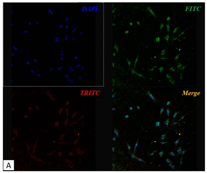
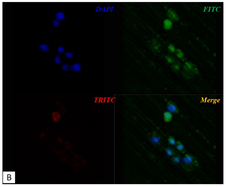
Figure 3.
Confocal microscope images: MLO-Y4 cell growth on treated plates (A) shows the dendritic shape characteristic of osteocyte, meanwhile cells maintain a rounder shape when grown on untreated surface (B). DAPI in figure marks nuclei in blue, FITC marks vitronectin in green, TRITC marks fibronectin in red. Panel A: 1000×, panel B: 4000×.


