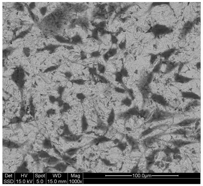
Figure 4.
MLO-Y4 under scanning electron microscope (SEM): Characteristic dendritic shape of MLO-Y4 cultured on treated titanium plates is also appreciable under SEM analysis where cell bodies and cytoplasmic processes (dendrites) are easily identifiable thanks to the adjustable gray contrast resulting from the analysis after the gold-palladium sputtering of samples. Magnifications and scale are reported in the figure.

