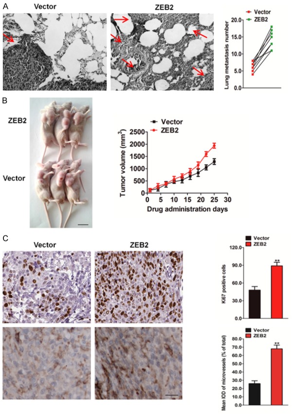
Figure 6.

Over-expression of ZEB2 contributes to HCT116 cells growth and metastasis in vivo. A. Metastatic lesions in the lungs of the mice which injected with HCT116-ZEB2 cells or control cells via tail vein (left). The total numbers of lung metastatic lesions in the ZEB2 transfection groups were much higher than those in controls (right). Scale bar represents 100 μm. B. The tumor growth was significantly promoted in mice subcutaneously inoculation with HCT116-ZEB2 cells versus control. Representative images of tumor-bearing mice (left) and tumor volumes were measured on the indicated days (right). Scale bar represents 1 cm. C. Mice bearing HCT116 tumor xenograft were sacrificed at the end of the experiment, and tumor tissues were removed for immunohistochemistry analysis with anti-Ki67 and anti-CD31 antibodies. Immunohistochemistry staining demonstrated the expression of Ki67 and CD31 positive cells in the indicated tissues. The data were presented as the mean ± SD. *P < 0.05, **P < 0.01 vs. vector group.
