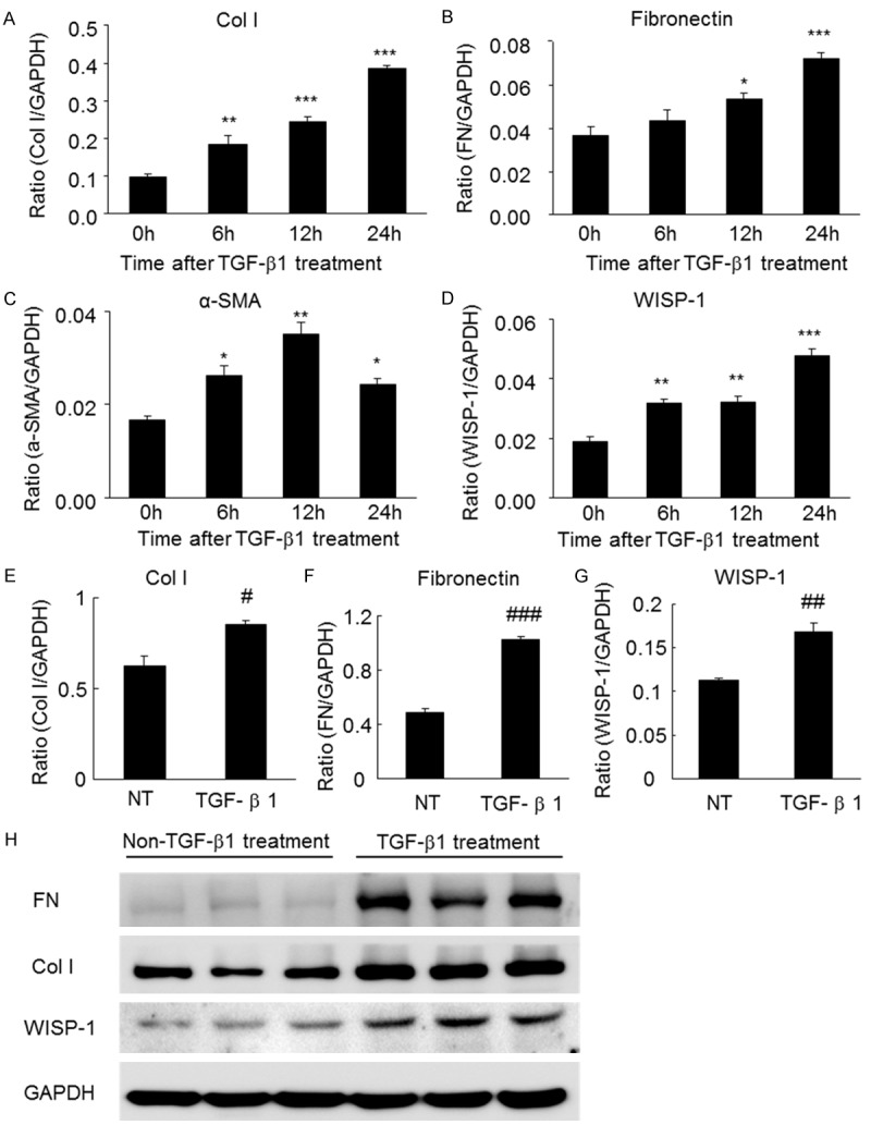
Figure 1.

The levels of WISP-1 and fibrosis markers in TGF-β-treated TECs were assessed using real-time PCR and western blot analyses. (A) The levels of collagen I, (B) fibronectin, (C) α-SMA and (D) WISP-1 are increased in TECs in response to the TGF-β treatment (2 ng/ml for 24 hours), as determined by real-time PCR analyses. (E) The protein levels of collagen I, (F) fibronectin and (G) WISP-1 are increased in TECs in response to the TGF-β treatment (2 ng/ml for 24 hours), as determined by western blot analyses. (H) Western blots of collagen I, fibronectin, α-SMA and WISP-1 in TECs treated with or without TGF-β (2 ng/ml for 24 hours). α-SMA, α-smooth muscle actin; Col I, collagen I; FN, fibronectin; NT, not treated with TGF-β; TGF, transforming growth factor; TECs, tubular epithelial cells. *P<0.05, **P<0.01, ***P<0.001 compared with 0 h. #P<0.05, ##P<0.01, ###P<0.001 compared with NT.
