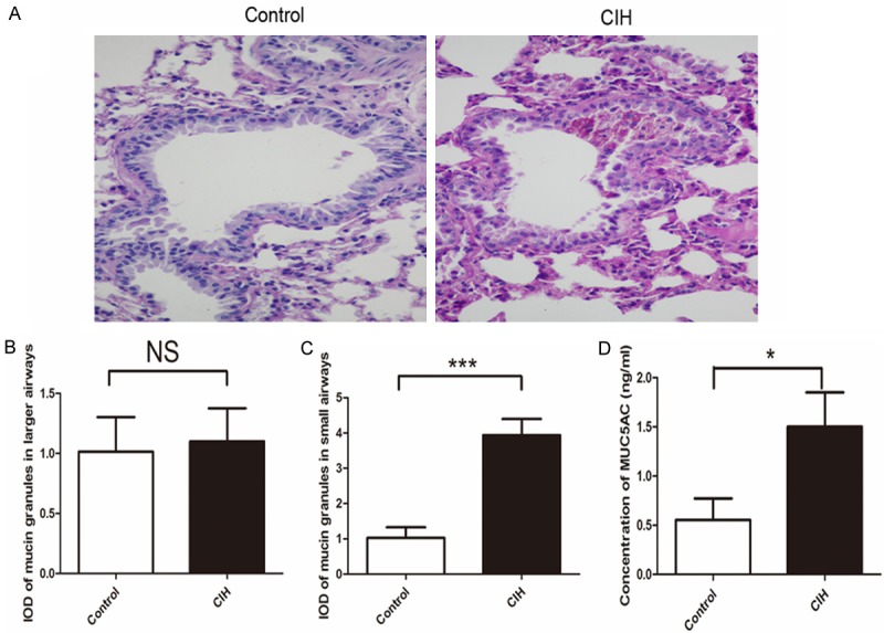
Figure 5.

Mucus secretion in mice was analyzed by periodic acid-Schiff (PAS) staining in the lung in these two groups. The left lung lobes were stained with PAS staining. The slides were scanedati 400 × magnifications (A), and the integrated optical density of mucin granules in large (B) and small airways (C), double-blind analysis performed in Image-Pro Plus 6.0. (D) Enzyme-linked immunosorbent assay results of MUC5AC in bronchoalveolar lavage fluid. Concentration of MUC5AC protein in the bronchoalveolar lavage fluid calculated from the standard curve in the same experiments (n=6 per group, P<0.05). Data were given as mean ± standard deviation (n=8), *P<0.05, **P<0.01, ***P<0.001. Data represent three independent experiments. All data were analyzed by SPSS using independent-sample t-test.
