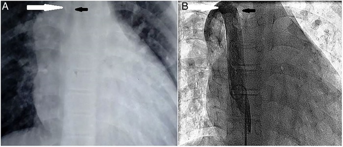
Figure 2.
Comparison of the chest X-ray film and the fluoroscopic image. (A) Chest X-ray, posteroanterior view, showing a smooth mass (white arrow) to the right of right tracheobronchial angle (black arrow). (B) Fluoroscopic image of venogram taken through a pigtail catheter placed in the dilated azygos vein. White arrow represents the dilated azygos vein entering the superior vena cava, corresponding to the smooth mass on chest X-ray while the black arrow corresponds to the right tracheobronchial angle.

