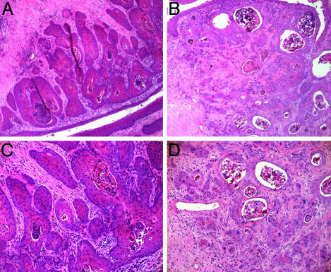
Fig. 4.
Cancer phenotype alterations in mice withdrawn from exogenous estrogen treatment. Shown are H&E-stained histological cross sections of tumors arising in the K14E6E7 mice after differing estrogen treatments. Withdrawal of exogenous estrogen led to a much more organized phenotype in the tumor tissue (A) than in mice given 9 months of continuous estrogen treatment (B). Notice the more organized structure in A, where most masses are confined by a basement membrane. Additionally, B shows more fibrous stromal reaction that is indicative of malignancy. At higher magnification, mice withdrawn from exogenous estrogen treatment (C) show less nuclear atypia and a lower number of individually invasive cells than mice given 9 months of continuous estrogen treatment (D).

