Figure 7.
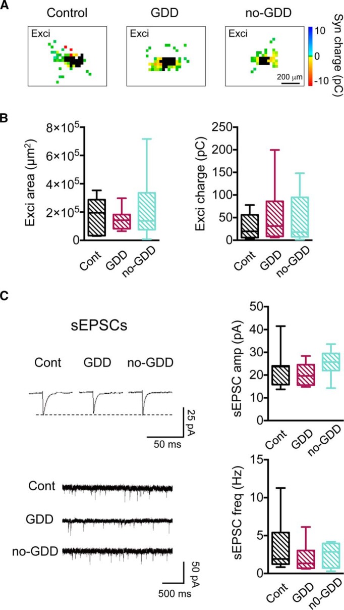
Synaptic inputs onto type 2 GABAergic neurons are unchanged in noise-traumatized mice. A, Examples of excitatory input maps of type 2 GABAergic neurons from control, GDD, and no-GDD mice. Uncaging sites that elicited direct responses in the recorded neuron are marked in black. B, Noise exposure had no effect on excitatory input area (left) or on total excitatory postsynaptic charge (right; area: control, n = 9 neurons, n = 8 animals, GDD, n = 7 neurons, n = 4 animals, no-GDD, n = 10 neurons, n = 6 animals, H = 0.067, p = 0.97, Kruskal–Wallis test; charge: control, n = 9 neurons, n = 8 animals, GDD, n = 7 neurons, n = 4 animals, no-GDD, n = 10 neurons, n = 6 animals, F(2,23) = 0.49, p = 0.62, one-way ANOVA). C, Amplitudes (top) and frequency of spontaneous EPSCs are unchanged by noise trauma (control, n = 7 neurons, n = 6 animals, GDD, n = 6 neurons, n = 3 animals, no-GDD, n = 10 neurons, n = 6 animals, amplitudes: F(2,20) = 1.13, p = 0.34, one-way ANOVA; frequency: F(2,20) = 1.00, p = 0.38, one-way ANOVA). Data are shown as box-and-whisker plots. *p < 0.05, **p < 0.01 in post hoc, pairwise assessments corrected for multiple comparisons.

