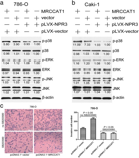
Fig. 7.

MRCCAT1 suppresses NPR3 to potentiate p38- MAPK signaling. a Western blot analysis of indicated proteins in MRCCAT1 overexpressed and control 786-O cells transfected with pLVX -NPR3. β-actin was used as an internal control. b Western blot analysis of indicated proteins in MRCCAT1 overexpressed and control Caki-1 cells transfected with pLVX -NPR3. β-actin was used as an internal control. c Left: Transwell assays in MRCCAT1 overexpressed and control 786-O cells treated with SB203580. Scale bar = 200 μm. Right: The statistical graph indicates the number of cells averaged from 8 random high power fields. The results are presented as the mean ± SD from three independent experiments. P values were calculated by Student’s t-test
