Fig. 1.
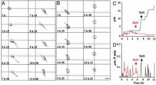
Time-lapse videomicroscopy recording of cerebellar granule cells cultured during 12 h in control conditions (A, C, and D)orinthe presence of C2-ceramide (B–D). (A and B) Microphotographs illustrating time-dependent changes of the cell morphology and motility in control conditions (A) or in the presence of 20 μM C2-ceramide (B). (C and D) Diagrams showing the distance covered by the recorded cells (C) and their velocity (D) in control conditions (black curves) or in the presence of 20 μM C2-ceramide (red curves). Scale bar, 8 μm.

