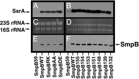
Fig. 3.
Ribosome association. (A and B) Northern blot analysis using an SsrA-specific probe to detect SsrA RNA in purified ribosome preparations. (C and D) Ethidium bromide staining of the same gel as in A and B, shown to demonstrate that similar amounts of ribosomal RNA were loaded in each lane. (E and F) Western blot analysis using anti-his6 antibody to detect his6-tagged SmpB protein in the same purified ribosome preparations used in A–D. The SmpB variant expressed in the cells from which the ribosomes were purified is indicated on the horizontal axis.

