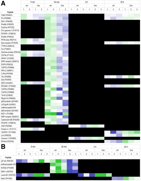
Fig. 6.

Protein tyrosine kinase (PTK, panel a) and serine/threonine kinase (STK, panel b) activity profiles at 5 min, 30 min, 2 h and 24 h in A7r5-α2Bvascular smooth muscle cells treated with 100 nM dexmedetomidine (DEX) or vehicle (ctrl). Peptides on the chips represent known phosphorylation sites of PTKs and STKs; for clarity, the names of these kinases and their UniProt identifiers are used instead of the peptide sequences. Two artificial (ART025 and ART004) peptides are included in the figure; they are not derived from natural proteins, but they were selected from a library of peptides known to be targets of kinase phosphorylation. Green and blue colors indicate increased or decreased kinase activity, respectively, in dexmedetomidine-treated cells when compared with vehicle-treated controls. Black color indicates no differences between dexmedetomidine-treated cells and vehicle-treated controls. The replication CV of high signal spots on the PTK and STK chips was <20%, indicating good experimental quality. n = 3 per time point; except for t = 5 min in the PTK experiment, t = 30 min (ctrl) and t = 24 h (ctrl) in the STK experiment, where n = 2
