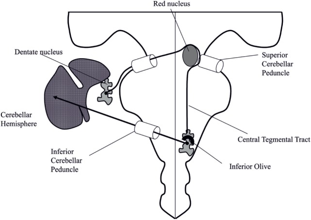
Figure 2.

Schematic representation of the Guillain–Mollaret triangle. The pathway coming from the contralateral dentate nucleus, through the contralateral brachium conjunctivum crosses the midline, turns around the ipsilateral red nucleus, and descends in the ipsilateral central tegmental tract to the inferior olive. Adapted from Ref. (36).
