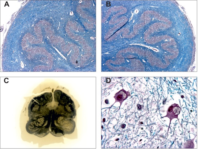
Figure 3.
Pathological features of degenerative inferior olive hypertrophy. Hypertrophic inferior olive (A) compared to contralateral side (B) (Bodian Luxol, X200). Note the mild demyelination of the surrounding white matter. (C) Coronal section of the medulla oblongata showing hypertrophy of the left inferior olive (Loyez stain). (D) Swelled and vacuolated nerve cells (“fenestrated neurons”) observed in the hypertrophic inferior olive [from (A), Bodian Luxol, X400]. Courtesy of Charles Duyckaerts and Franck Bielle, Escourolle’s Lab, Pitie-Salpetriere Hospital, Paris, France. Adapted from Ref. (51).

