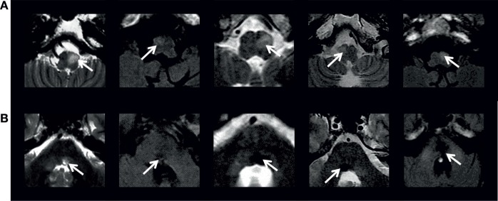
Figure 4.
Axial FLAIR or T2 MRI (1.5-T GE scanners) at (A) inferior olive level and (B) midpontine tegmentum level in five patients with symptomatic oculopalatal tremor. White arrows in (A) indicate the abnormal inferior olive hypersignal and in (B) the causative lesion. Adapted from Ref. (24).

